Labeled Small Intestine Cell Diagram

Clinical significance of small intestine.
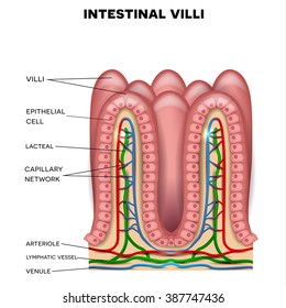
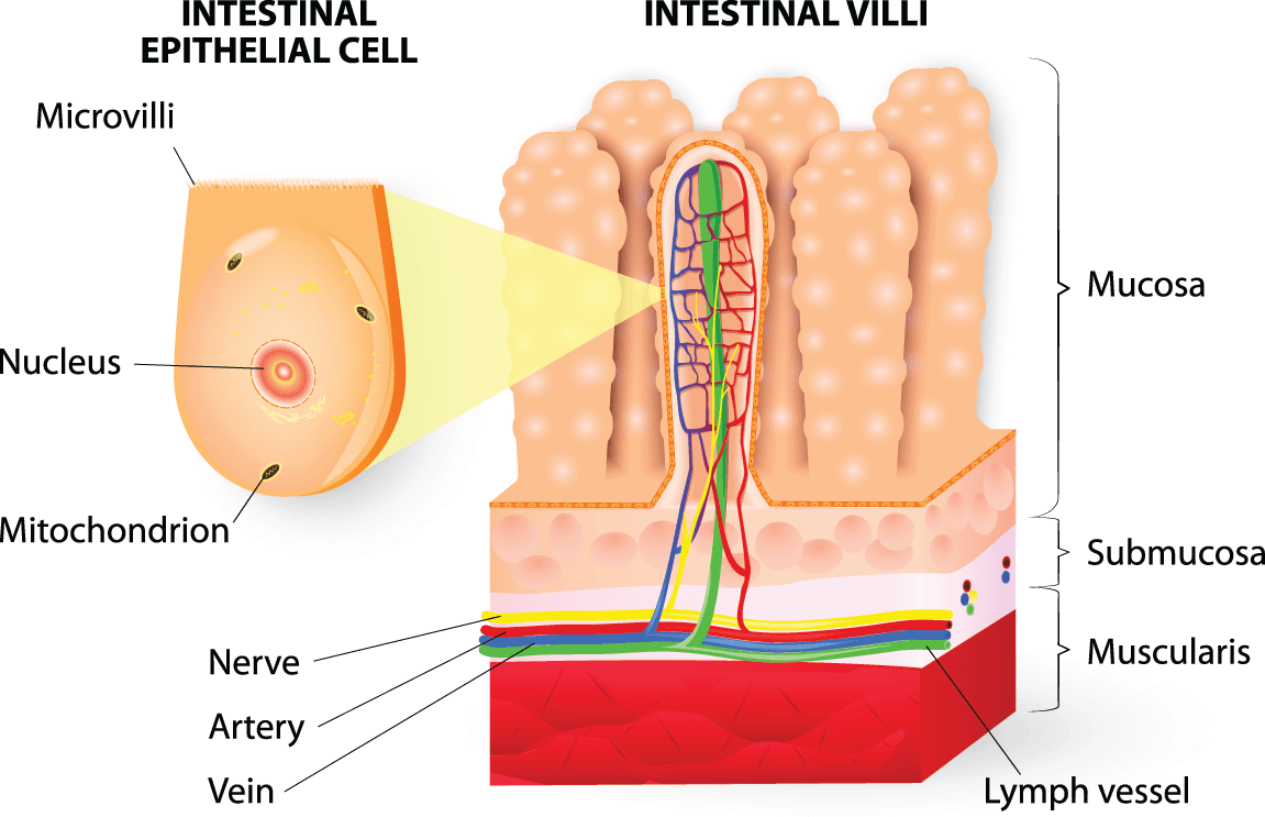
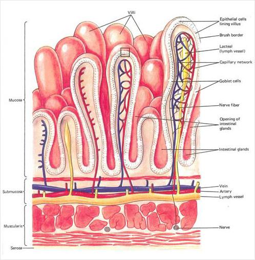
Labeled small intestine cell diagram. Learn about its parts location in the body function and conditions that affect the intestines. Compare and contrast the location and gross anatomy of the small and large intestines. This distinguishes the small intestine from the stomach. The large intestine part of the human digestive system.
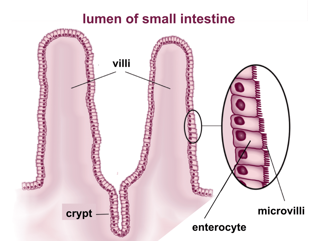
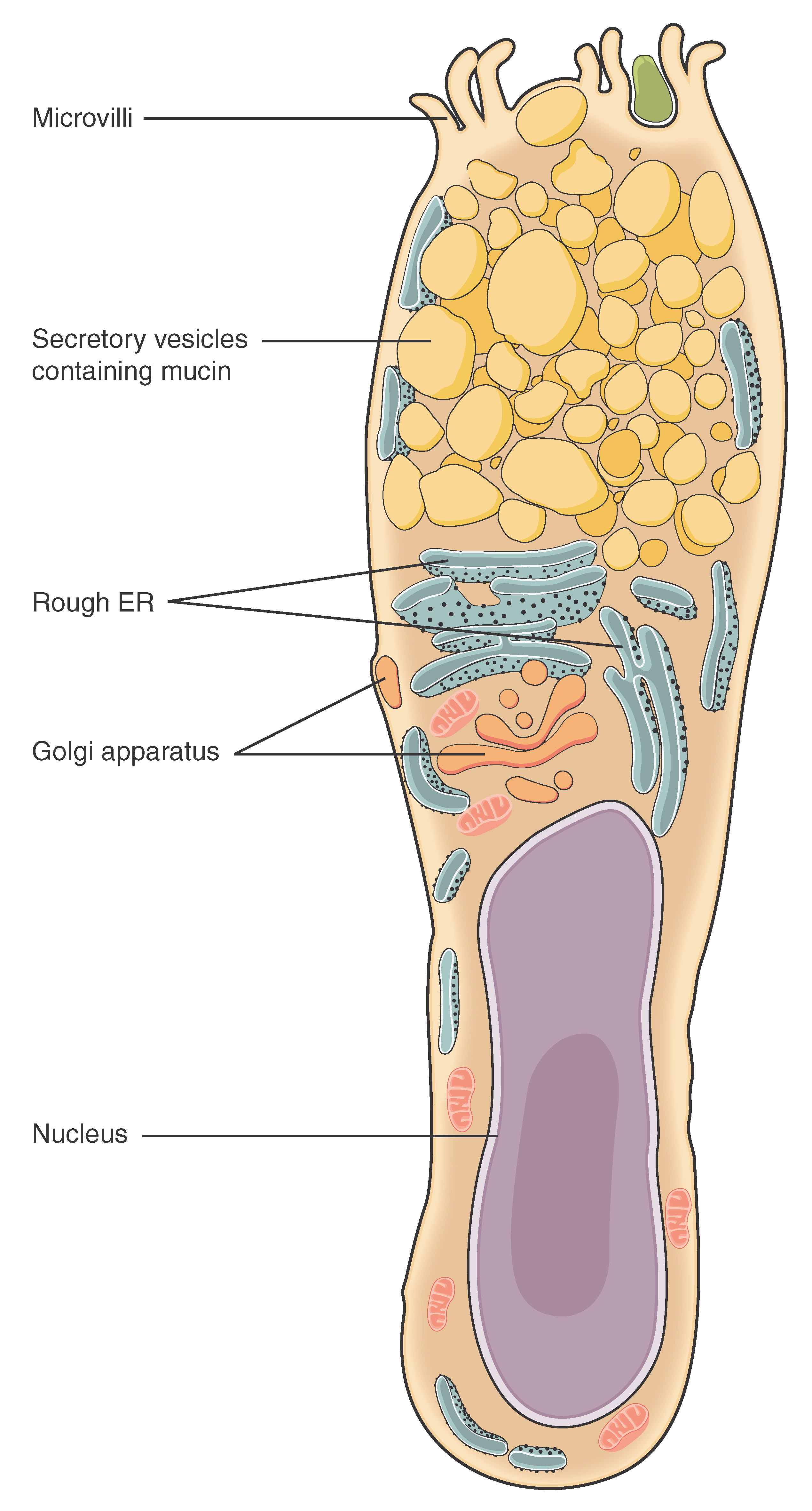
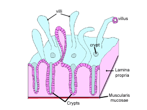
Thousands of microvilli are present on the apical surface of the epithelial cells in the small intestine. We are pleased to provide you with the picture named three parts of small intestine diagramwe hope this picture three parts of small intestine diagram can help you study and research. In living humans the small. The small intestine is a organ located in the gastrointestinal tract which assists in the digestion and absorption of ingested food.
The immunological cells attack the small intestine thereby. The duodenum jejunum and ileum. A number of clinical conditions harm the small intestine. Learn vocabulary terms and more with flashcards games and other study tools.
It occurs when the absorptive cells of the small intestine do not produce enough lactase the enzyme that digests the milk sugar lactose. By the end of this section you will be able to. The small and large intestines. Start studying anatomy of small intestine.
Large labelled diagram of the anatomy of large intestine including the main structure of the large intestine. That is enzymatic digestion occurs not only in the lumen but also on the luminal surfaces of the mucosal cells. This introductory level educational material is suitable for high school students gcse as a2 a level itec and students of first level health sciences subjects including diet and nutrition. These form a brush border.
Simple columnar cells in the small intestine have several tiny hair like surface projections called microvilli. These are tightly packed and their length might be anywhere between 05 to 10 mm. It extends from the pylorus of the stomach to the iloececal junction where it meets the large intestine. The small intestine is made up of the duodenum jejunum and ileum.
Small intestine location and anatomy. Anatomically the small bowel can be divided into three parts. Together with the esophagus large intestine and the stomach it forms the gastrointestinal tract. Starting from physical obstruction of the lumen to bacterial or viral symptoms there are many complex disorders.

